Biomedical Imaging Core
The biomedical imaging core supports department investigators by providing training and access to cutting edge optical imaging platforms. All of these resources require one or more training sessions before use, and some of them require scheduling. Please contact Dr. Cojen Ho if you wish to use these resources.
Nikon Widefield Live Cell Imaging Rig
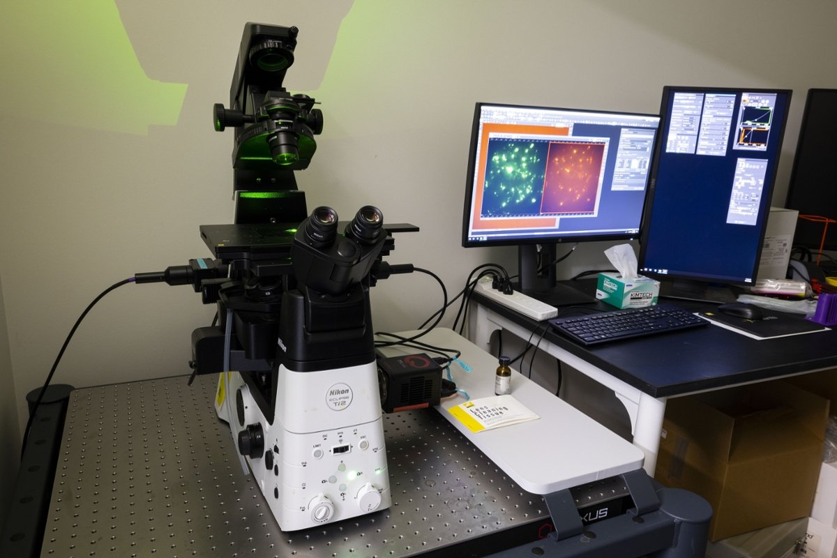 This microscope is useful for calcium, biosensor/GFP and FRET imaging. It can also be used for standard widefield microscopy of fluorescent samples. It is located in the Joint Health Sciences Center. There is an online calendar for reservations.
Technical Specifications: Nikon Ti2 inverted microscope equipped with 20, 60, and 100X Nikon SuperFluor objectives and DIC, an ORCA fusion sCMOS camera, and Nikon Perfect Focus System (PFS). The microscope is driven by Nikon Elements. Excitation and emission are controlled by Ludl filter wheels.
Nikon Total Internal Reflection Fluorescence (TIRF) Microscopy Rig
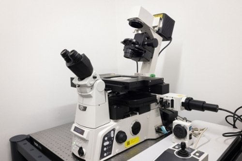 This microscope is used for single particle tracking and/or live cell imaging of near plasma membrane events in cells expressing fluorescent proteins. It is located in the Joint Health Sciences Center.
Technical Specifications: Nikon Ti inverted TIRF microscope equipped with 60X Apo TIRF oil objective, a Photometrics Prime 95B sCMOS camera, laser launch with 488 and 561 laser lines, and NIS-Elements control and acquisition software. This purpose-built TIRF rig is also equipped with the Nikon Perfect Focus System (PFS). There is also a separate dedicated workstation for data analysis of TIRF images offline.
Nikon Inverted Confocal Microscope
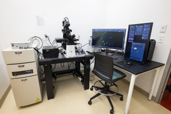 This is an inverted laser scanning confocal microscope which is equipped with a resonant scanner for high speed imaging and artificial intelligence (AI)-based image processing and deconvolution. This microscope can be used for both live cell and fixed sample imaging. There is a dedicated imaging suite for this microscope in the Medical School Education (MSE) building. There is an online calendar for reservations.
Technical Specifications: Nikon A1R HD25 25mm field of view inverted confocal microscope with 4 laser lines and a high definition resonant scanner capable of 1024x1024 resolution or up to 720FPS. Image detection is through a 4 channel detector with 2 GaAsP and 2 high sensitivity PMTs.
Nikon Upright Spinning Disk Confocal Microscope
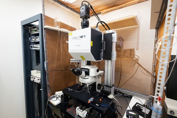 This is an upright Yokogawa spinning disk ultra-widefield confocal microscope which can also support simultaneous electrophysiological recording. There is a dedicated imaging suite for this microscope is located in the Medical School Education (MSE) building.
Technical Specifications: Nikon CSU-W1 spinning disk upright confocal with 2 laser lines. Image detection is with an ORCA-FusionBT back-thinned camera. This rig is also equipped for electrophysiological recordings.
ONI Nanoimager Superresolution Microscope
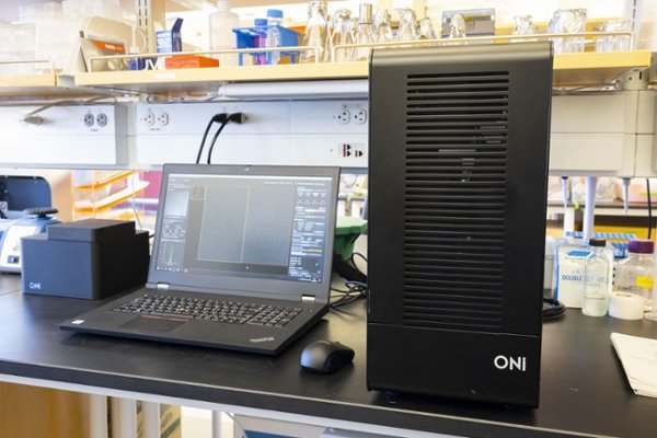 This is a multi-modal imaging platform which support both live cell and fixed sample imaging using PALM and dSTORM superresolution techniques. It can also perform widefield and TIRF imaging. This microscope is located in the Medical School Education (MSE) building.
Technical Specifications: ONI Nanoimager with 100X objective, 4 laser lines, 2D and 3D PALM, 2D and 3D dSTORM, single particle tracking, and single molecule FRET. Three different illumination modes include widefield, TIRF, and HILO. NimOS software includes both acquisition and analysis features.
Abberior STEDYCON Superresolution STED and Confocal Microscope
This is a stimulated emission depletion (STED) superresolution
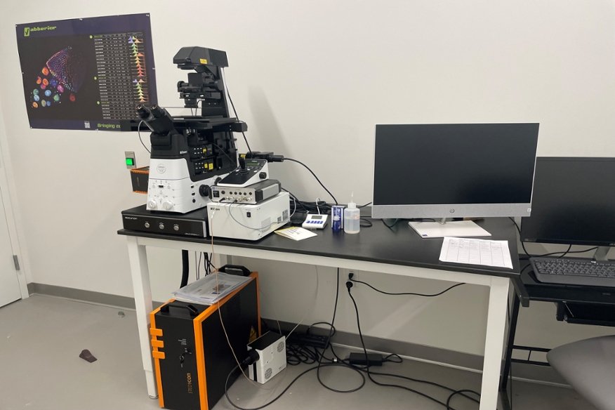
microscope which can also perform traditional laser scanning confocal imaging. It is located in the Joint Health Sciences Center.
Technical Specifications: Abberior STEDYCON superresolution STED/confocal microscope with four confocal laser lines. The STED laser is a 775nm pulsed laser. This superresolution microscope has two STED and four confocal imaging channels. Detection is with four single photon counting avalanche photodiode (APD) modules.