Mitochondrial Physiology Core
The mitochondrial physiology core supports department investigators by providing training and access to metabolic profiling instruments. These resources require one or more training sessions before use. Please contact Dr. Kleiton Silva or Dr. Darren Boehning prior to using these resources.
Agilent Seahorse XFe96
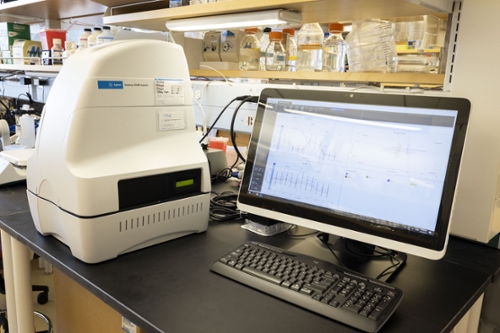 There is an Agilent Seahorse XFe96 Analyzer available for oxygen consumption rate and proton efflux rate in mitochondria in a 96 well format. This instrument can measure mitochondrial function in isolated mitochondria, cultured cells, or tissue fragments. Please contact Dr. Kleiton Silva regarding training, use, and availability. This instrument is located in the Medical School Education (MSE) building.
Technical Specifications: 96 well capacity, precision temperature control, semi-automated acquisition of metabolic data in real time. Analysis of as few as 5000 cells/well. Includes four injection ports with automated mixing to measure the mitochondrial response to compounds in real time in live cells. Wave software is used for protocol creation and data analysis.
Molecular Devices FlexStation3 Multi-mode Plate Reader
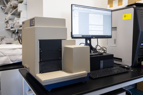 Molecular Devices FlexStation 3 multi-mode plate reader is available for semi-automated measurement of mitochondrial polarization using fluorescent dyes. Multiple injections of compounds is possible using robotic liquid handling in a high throughput format (up to 384 wells). In addition to measuring mitochondrial polarization, this plate reader is the industry standard reader for calcium fluxes, enzyme assays, and reporter assays, and is widely used by the calcium imaging field. This instrument is located in the Joint Health Sciences Center.
Technical specifications: Xenon flash illumination with monochromator-based filters tunable in 1.0 nm increments. Capable of imaging in 6 through 384 well formats with endpoint, kinetic, spectral scanning, and well scanning modes. Detection is mediated by two photomultiplier tubes (PMTs). Our optioned model does absorbance, fluorescence, luminescence, fluorescence polarization, and homogeneous time resolved fluorescence.